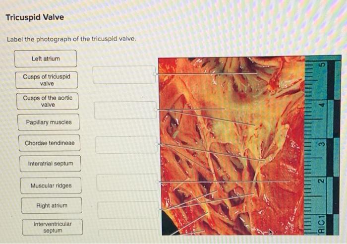
Label the photograph of the tricuspid valve to delve into the intricate anatomy of this crucial cardiac structure. Understanding its key components and function is essential for comprehending the complexities of the heart’s circulatory system.
This comprehensive guide provides a detailed overview of the tricuspid valve, its role in the cardiac cycle, and the implications of its dysfunction. Discover the importance of this valve in maintaining unidirectional blood flow and preventing regurgitation.
1. Label the Photograph of the Tricuspid Valve
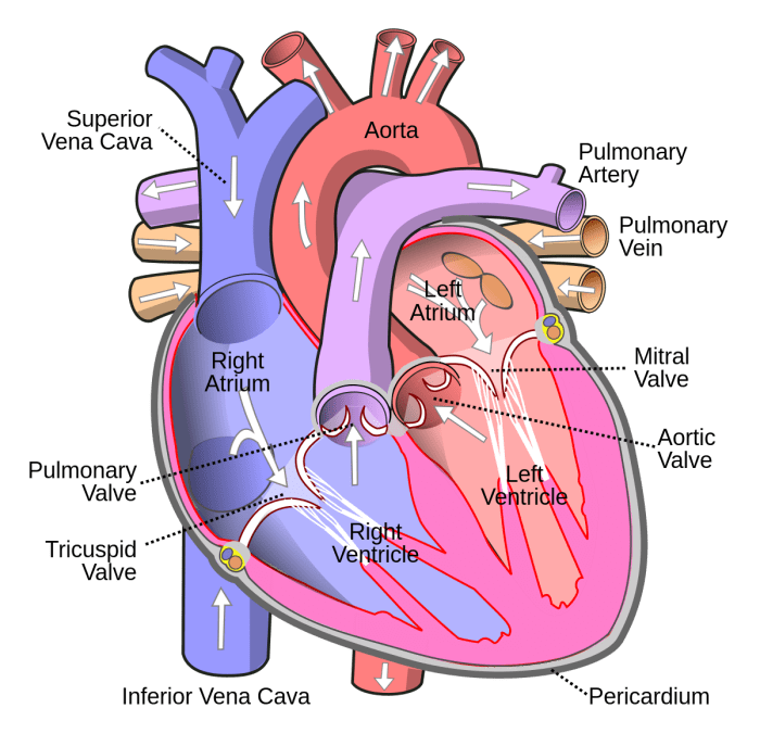
The tricuspid valve is a heart valve located between the right atrium and the right ventricle. It is composed of three cusps or leaflets that open and close to allow blood to flow from the atrium to the ventricle.
The photograph below shows a labeled photograph of the tricuspid valve. The key anatomical structures of the tricuspid valve are identified as follows:
- Anterior cusp:The anterior cusp is the largest of the three cusps. It is located on the front of the valve.
- Posterior cusp:The posterior cusp is located on the back of the valve.
- Septal cusp:The septal cusp is located between the anterior and posterior cusps. It is attached to the interventricular septum.
- Chordae tendineae:The chordae tendineae are thin, fibrous cords that attach the cusps to the papillary muscles of the ventricle. They prevent the cusps from prolapsing into the atrium when the valve is closed.
- Papillary muscles:The papillary muscles are small muscles located on the walls of the ventricle. They contract to pull the chordae tendineae and close the valve.
2. Function of the Tricuspid Valve

The tricuspid valve plays a critical role in the cardiac cycle. It opens during systole, the contraction phase of the heart, to allow blood to flow from the right atrium to the right ventricle. It closes during diastole, the relaxation phase of the heart, to prevent blood from flowing back into the atrium.
The tricuspid valve is essential for maintaining proper blood flow through the heart. If the valve is damaged or diseased, it can lead to tricuspid valve regurgitation, a condition in which blood leaks back into the atrium during systole. This can lead to heart failure and other serious complications.
3. Tricuspid Valve Disease: Label The Photograph Of The Tricuspid Valve
Tricuspid valve disease is a condition in which the tricuspid valve is damaged or diseased. This can lead to tricuspid valve regurgitation, a condition in which blood leaks back into the atrium during systole. Tricuspid valve disease can be caused by a variety of factors, including:
- Rheumatic fever
- Infective endocarditis
- Cardiomyopathy
- Congenital heart defects
The symptoms of tricuspid valve disease can vary depending on the severity of the condition. Some people may experience no symptoms, while others may experience:
- Shortness of breath
- Fatigue
- Swelling in the legs, ankles, and feet
- Abdominal pain
- Chest pain
Treatment for tricuspid valve disease depends on the severity of the condition. In some cases, no treatment is necessary. In other cases, treatment may include medications, surgery, or a combination of both.
4. Echocardiography of the Tricuspid Valve

Echocardiography is a non-invasive imaging technique that uses sound waves to create images of the heart. Echocardiography can be used to evaluate the structure and function of the tricuspid valve.
There are different echocardiographic views that can be used to assess the tricuspid valve. These views include:
- Apical four-chamber view:This view shows the four chambers of the heart, including the right atrium, right ventricle, left atrium, and left ventricle.
- Parasternal short-axis view:This view shows a cross-section of the heart at the level of the tricuspid valve.
- Subcostal view:This view shows the heart from below the rib cage.
Echocardiography can be used to diagnose tricuspid valve disease and to assess the severity of the condition. It can also be used to monitor the effectiveness of treatment.
5. Surgical Management of Tricuspid Valve Disease

Surgical intervention may be necessary for patients with severe tricuspid valve disease. Surgical options include:
- Tricuspid valve repair:Tricuspid valve repair involves repairing the damaged or diseased valve. This can be done through a variety of techniques, including:
- Annular plication:This technique involves tightening the ring around the valve to reduce leakage.
- Chordal replacement:This technique involves replacing the damaged chordae tendineae with artificial chords.
- Tricuspid valve replacement:Tricuspid valve replacement involves replacing the damaged or diseased valve with a mechanical or biological valve.
The choice of surgical intervention depends on the severity of the tricuspid valve disease and the patient’s overall health.
Query Resolution
What is the function of the tricuspid valve?
The tricuspid valve prevents backflow of blood from the right ventricle into the right atrium during ventricular contraction.
What are the common types of tricuspid valve disease?
Tricuspid valve disease includes regurgitation (leaky valve) and stenosis (narrowed valve).
How is tricuspid valve disease diagnosed?
Echocardiography is the primary imaging technique used to evaluate the tricuspid valve’s structure and function.